Advancing Surgical Frontiers: Exploring the Potential of Digital Innovations in Minimally Invasive Surgery

Before 8 o'clock in the morning, Professor Piet Pattyn arrived at Ghent University Hospital by bike from the south side. As soon as he entered the room, he first checked with his colleagues about the patients' conditions from the night before. The atmosphere was charged with purpose; three laparoscopic surgeries awaited him, each a testament to his skill and dedication.
This year marks Professor PattynˇŻs 40th at Ghent University Hospital, one of Belgium's largest medical centers. He has dedicated his career to gastrointestinal surgery, witnessing the shift from open procedures to minimally invasive techniques and the continuous advancement of surgical technology.

ˇ°The early stages of minimally invasive instruments were reminiscent of the first Macintosh computersˇŞblurry displays and unresponsive controls. Back then, the risks associated with surgery were also higher due to technological limitations,ˇ± said Professor Pattyn. ˇ°However, advancements in technology have injected a steady stream of energy into our work, and I can truly feel this transformation.ˇ±
This summer, the 32nd Annual International Conference of the European Association for Endoscopic Surgery (EAES) took place in Maastricht, Netherlands. We seized the opportunity to gather with leading surgical experts and explore the transformative impact of digital intelligence on minimally invasive surgery.
Enhancing Surgeons' Vision: Clearer and Broader Perspectives
Professor Pattyn recalls that when they began experimenting with gastrointestinal minimally invasive surgery in the 1990s, the lack of high-definition imaging systems posed significant challenges in visualizing the surgical field. The issue of obtaining clear images consistently became a common concern among surgeons.
Fr¨¦d¨¦ric De Ryck, a cardiothoracic surgeon at Ghent University Hospital, shares similar sentiments. After recently completing a Single-port thoracoscopic lobectomy and lymphadenectomy, he expressed how Mindray's new-generation endoscopic system, HyPixel UX5, left a lasting impression on him.
ˇ°Image clarity is crucial in minimally invasive surgery. Today, I used a 5mm laparoscopy and related endoscopic systems, and the 4K images with ultra-high color reproduction greatly enhanced the ability to visualize the surgical field.ˇ±
In addition to ultra-high-definition imaging, Professor Pattyn focuses on 3D imaging technology as a gastrointestinal surgery expert. ˇ°Especially during suturing or complex resections, a 3D view allows me to perceive the three-dimensional structure of the organs more clearly,ˇ± he remarked. ˇ°Even though IˇŻm operating through small incisions, the immersive experience feels akin to performing open surgery.ˇ±
Moreover, the application of fluorescence technology has emerged as another hot topic in the industry. Many experts believe that fluorescence can significantly enhance surgical precision, leading to better postoperative recovery for patients. Professor Mario Annecchiarico elaborated on this based on his own experiences.

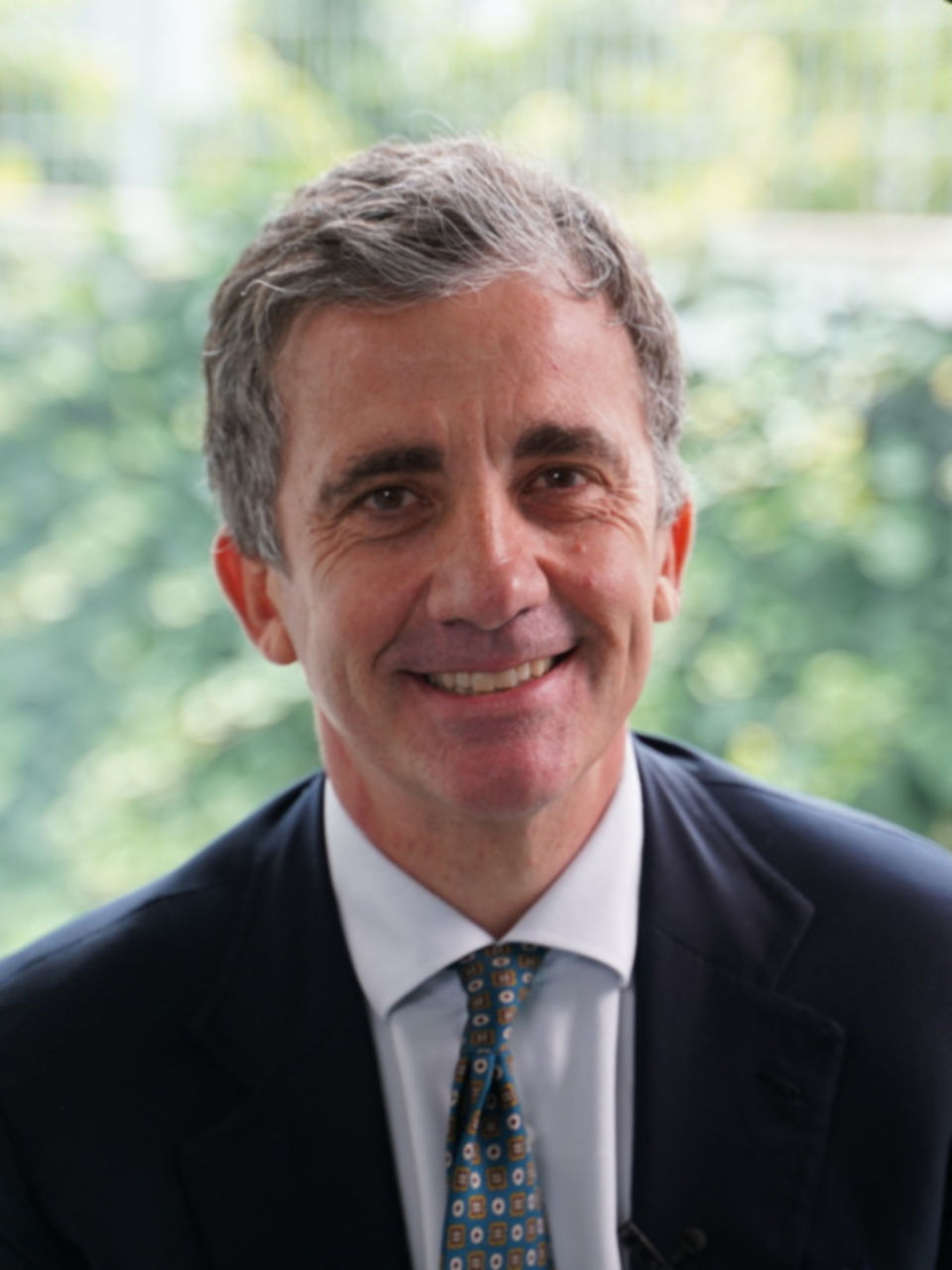
Prof. Mario Annecchiarico
Director of General Surgery Unit, Major National Hospital "San Pio" of Benevento
I often use fluorescence during surgeries, particularly for colorectal surgery and at the gastroesophageal junction. It effectively helps me assess blood supply, which is crucial for reducing the risk of anastomotic leaks. Additionally, in the conversion treatment of locally advanced gastric cancer, utilizing fluorescence-guided techniques for navigating lymphadenectomy around the stomach allows for the resection of more lymph nodes and aids in the precise removal of the primary tumor.
Patient Safety is Always the Top Priority
ˇ°Patient safetyˇ± was the keyword most frequently discussed among the doctors we spoke withˇŞhow to employ innovative smart technologies to conduct safer and more efficient minimally invasive surgeries is every surgeonˇŻs ultimate pursuit.
ˇ°Today, I operated on a patient who had undergone an esophagectomy about 15 years ago. The structure of his stomach is quite weak, and there are adhesions present,ˇ± Professor Pattyn explained. ˇ°The ultrasonic scalpel used in todayˇŻs surgery made a deep impression on me. Its refined jaw design and comfortable grip made it much easier for me to dissect tissue adhesions. This was my biggest feeling from the surgery.ˇ±
At the same time, Professor Pattyn noted that surgeons have different preferences for energy devices based on their techniques and habits, yet the safety of the operation remains paramount.
MindrayˇŻs EVS enhanced vessel sealing algorithm accurately adjusts energy output based on the condition of the vessel, ensuring secure vessel coagulation during surgery and facilitating good postoperative recovery for the patient.
Professor Dario Scala from Italy advocates for the digitization of surgical procedures and holds high expectations for AI applications in minimally invasive surgery.
Dr. Dario Scala
Director of Department of Surgery in Caserta Hospital Sant 'Anna e San Sebastiano
Right now, artificial intelligence is a hot topic in the minimally invasive surgery field. I believe AI has the potential to enhance endoscopic imaging, giving doctors higher precision and resolution in tissue visualization. It can also help reduce distractions from smoke and steam during procedures, leading to more precise surgeries and ultimately better recovery outcomes for patients.
The Future of Smart Integration in Minimally Invasive Surgery
Beyond ˇ°clear visibility and precise cuttingˇ± many experienced surgeons share the vision of utilizing smart integration to elevate surgical techniques to new heights.
ˇ°If the anesthesia, patient monitor, and other vital signs could be displayed in real-time alongside endoscopic images, our control during surgery would become much more precise, greatly benefiting the development of minimally invasive surgery,ˇ± said Umberto Bracale, a professor in the medical department at the University of Salerno in Italy. He is particularly interested in the interconnection between endoscopic systems and other operating room equipments.
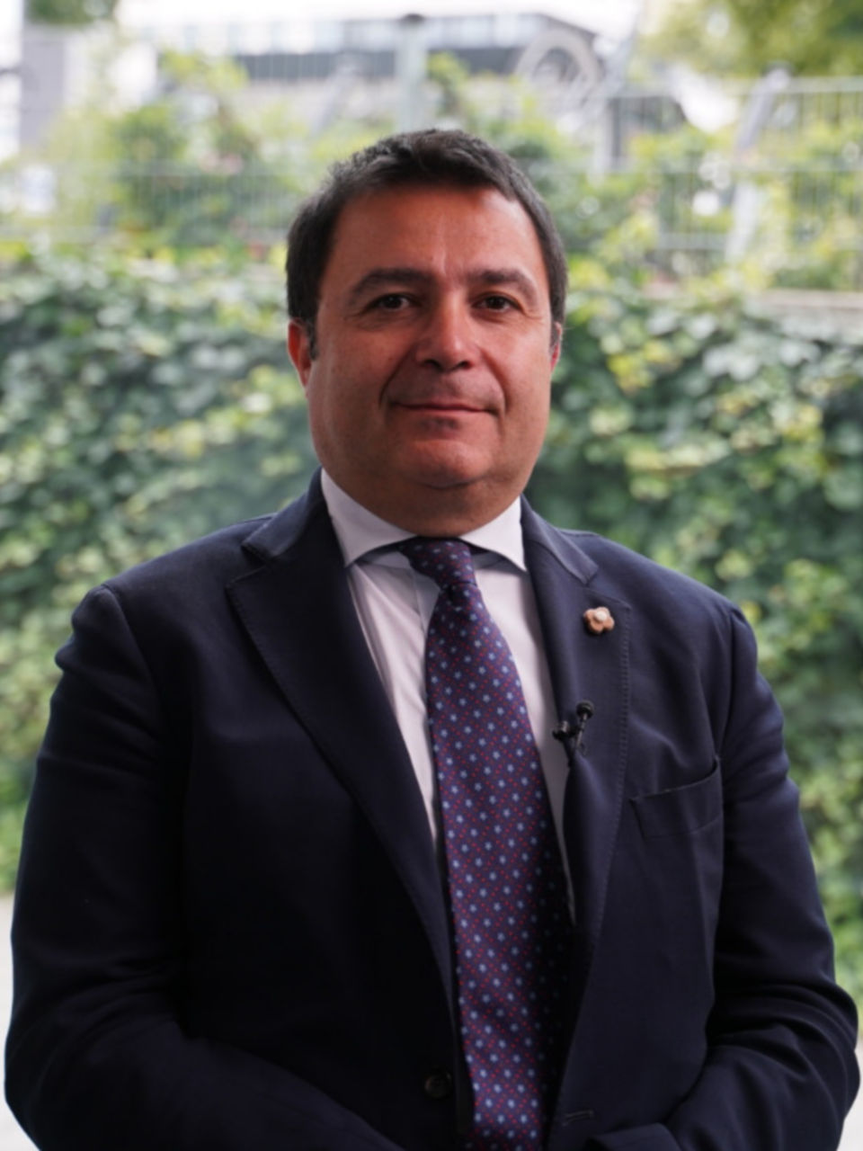
Prof. Umberto Bracale
Department of Medicine, Surgery and Dentistry University of Salerno ltaly
In liver resection surgery, the combination of ultrasound and endoscope camera system allows surgeons to see not just the external appearance of the organs but also the internal lesions and structural information. What's even better is that having both ultrasound and endoscopic images displayed on the same screen means surgeons donˇŻt have to switch back and forth between two machines. This makes our work a lot easier.
Regarding combined imaging, thoracic surgeon De Ryck believes this technology should surpass mere integration of ultrasound and endoscopic imaging. ˇ°Due to the unique softness of lung tissue compared to other organs, its inflation patterns both pre- and intraoperatively influence a surgeonˇŻs judgment regarding vessel locations. Therefore, real-time display of CT 3D reconstructed images of the lungs during surgery becomes essential.ˇ±

Bringing Advanced Technology to the World
"About ten years ago, I took part in a training program at a hospital in Shanghai," Dr. De Ryck recalled, reflecting on his experience in China. "I was truly impressed by the volume of surgeries performed by the medical staff there. In just two weeks, I observed more surgeries than I would typically see over a six-month period."
Like Dr. De Ryck, Professor Pattyn, who has also participated in international surgical initiatives, believes that global collaboration and exchange are crucial for advancing the field of minimally invasive surgery. The ultimate aim of all surgeons and healthcare technology companies is to deliver better recovery outcomes for patients through more precise and safer surgical techniques that are widely accessible.
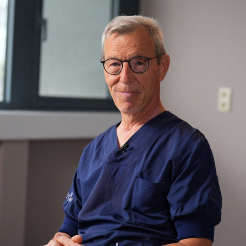
Prof. Piet Pattyn
Head of Gastrointestinal Surgery Department, Ghent University Hospital
Yesterday, I got a birthday card from the family of a patient I treated 20 years ago. It really warms my heart to know that someone I once cared for is now living healthily. ThatˇŻs what itˇŻs all about for me and what IˇŻve dedicated my life to. I always make sure to share this mindset with my students too.
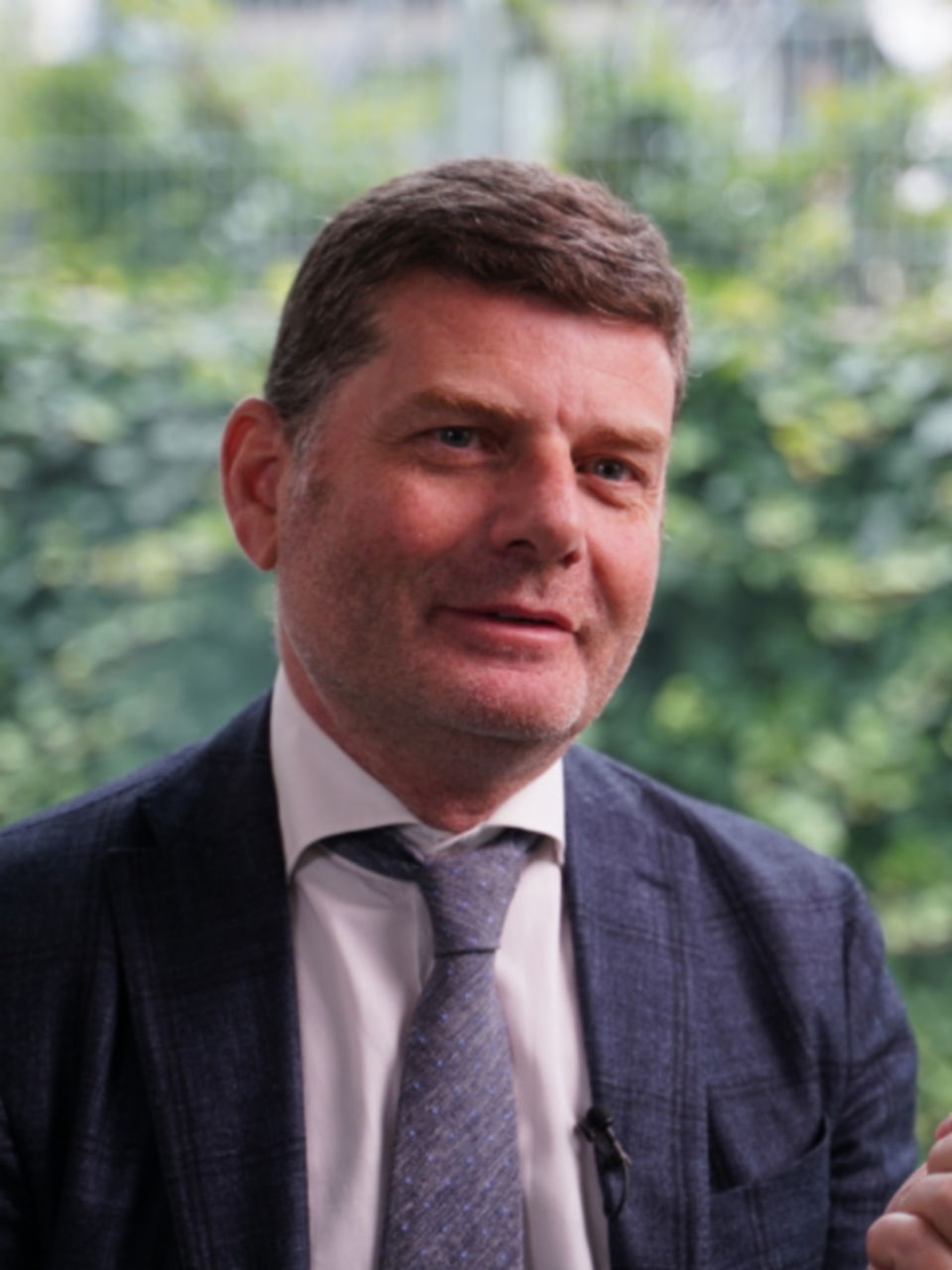
Dr. Fr¨¦d¨¦ric De Ryck
Vascular and Thoracic surgeon, Ghent University Hospital
As a doctor, I believe that no matter where we come from, we all share the same goal and belief: to perform more precise surgeries that minimize trauma for our patients. We want them to experience better treatment and recovery outcomes so they can get back to their healthy daily lives as soon as possible.
"In my view, every patient represents more than just an individual seeking treatment," Professor Pattyn noted. "They embody a family; they are husbands, wives, parents, or children."
